“Imaging Techniques for Bone Health Assessment
Related Articles Imaging Techniques for Bone Health Assessment
- Signs And Symptoms Of Bone Density Loss
- The Role Of Nutrition In Supporting Bone Density
- Bone Density Tests: Importance And Types
- The Role Of Hormones In Bone Growth And Development
- The Intricate Link Between Vitamin D Deficiency And Bone Health
Introduction
On this special occasion, we are happy to review interesting topics related to Imaging Techniques for Bone Health Assessment. Let’s knit interesting information and provide new insights to readers.
Absolutely! Here’s a comprehensive article on imaging techniques for bone health assessment, covering a wide range of methods, their principles, advantages, limitations, and clinical applications.

Imaging Techniques for Bone Health Assessment: A Comprehensive Overview
Introduction
Bone health is a critical aspect of overall well-being, playing a vital role in structural support, protection of internal organs, and mineral homeostasis. The assessment of bone health is essential for the diagnosis, monitoring, and management of various skeletal disorders, including osteoporosis, osteopenia, fractures, and metabolic bone diseases. Imaging techniques have revolutionized the field of bone health assessment, providing non-invasive or minimally invasive methods to evaluate bone density, structure, and integrity. This article provides a comprehensive overview of the various imaging modalities used in bone health assessment, discussing their principles, advantages, limitations, and clinical applications.
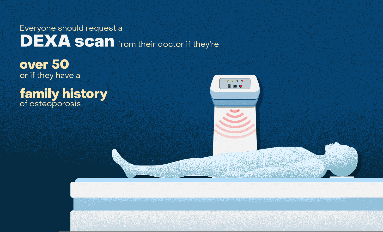
1. Dual-Energy X-ray Absorptiometry (DXA)
- Principle: DXA is the gold standard for measuring bone mineral density (BMD). It uses two X-ray beams with different energy levels to differentiate between bone and soft tissue. BMD is calculated as the area density of mineral in the bone (grams per square centimeter).
- Advantages:
- Low radiation dose.
- High precision and accuracy.
- Widely available and relatively inexpensive.
- Provides T-scores and Z-scores for comparison with reference populations.
- Limitations:
- Two-dimensional imaging, which does not provide information about bone microarchitecture.
- Can be affected by degenerative changes in the spine, such as osteophytes and vertebral compression fractures.
- May underestimate BMD in individuals with large body size.
- Clinical Applications:
- Diagnosis of osteoporosis and osteopenia.
- Monitoring response to osteoporosis treatment.
- Risk assessment for fractures.
- Assessment of BMD in various skeletal sites, including the spine, hip, and forearm.
2. Quantitative Computed Tomography (QCT)
- Principle: QCT uses computed tomography (CT) to measure BMD in three dimensions. It quantifies the volumetric bone density (milligrams per cubic centimeter) in trabecular and cortical bone.
- Advantages:
- Provides true volumetric BMD measurements, which are less affected by bone size and degenerative changes.
- Can assess BMD in specific regions of interest (ROIs) within the bone.
- Can be used to evaluate bone microarchitecture.
- Limitations:
- Higher radiation dose compared to DXA.
- More expensive than DXA.
- Requires specialized software and expertise.
- Clinical Applications:
- Assessment of BMD in individuals with spinal degeneration or large body size.
- Evaluation of bone microarchitecture.
- Research studies on bone metabolism and fracture risk.
3. High-Resolution Peripheral Quantitative Computed Tomography (HR-pQCT)
- Principle: HR-pQCT is a specialized CT technique that provides high-resolution images of bone microarchitecture in peripheral skeletal sites, such as the distal radius and tibia.
- Advantages:
- Non-invasive assessment of bone microarchitecture, including trabecular number, thickness, and separation.
- Provides information about cortical porosity and thickness.
- Can be used to predict fracture risk.
- Limitations:
- Limited to peripheral skeletal sites.
- Higher radiation dose compared to DXA.
- Requires specialized equipment and expertise.
- Clinical Applications:
- Research studies on bone microarchitecture and fracture risk.
- Evaluation of bone quality in individuals with metabolic bone diseases.
- Monitoring response to osteoporosis treatment.
4. Magnetic Resonance Imaging (MRI)
- Principle: MRI uses magnetic fields and radio waves to create detailed images of bone and soft tissues. It can assess bone marrow composition, trabecular structure, and bone perfusion.
- Advantages:
- No ionizing radiation.
- Excellent soft tissue contrast.
- Can detect bone marrow edema, which is a sign of bone stress or fracture.
- Can be used to evaluate bone tumors and infections.
- Limitations:
- More expensive than DXA and QCT.
- Longer scan times.
- Can be affected by metal implants.
- Not as sensitive as DXA for measuring BMD.
- Clinical Applications:
- Evaluation of bone marrow disorders.
- Assessment of trabecular structure.
- Detection of bone stress fractures.
- Diagnosis of bone tumors and infections.
5. Bone Scintigraphy (Bone Scan)
- Principle: Bone scintigraphy involves injecting a radioactive tracer into the bloodstream, which is then taken up by bone tissue. A gamma camera detects the tracer, creating an image of bone metabolism.
- Advantages:
- Highly sensitive for detecting areas of increased bone turnover.
- Can be used to evaluate the entire skeleton.
- Limitations:
- Low specificity, as increased bone turnover can be caused by various conditions.
- Radiation exposure.
- Limited information about bone structure.
- Clinical Applications:
- Detection of bone metastases.
- Evaluation of fractures and infections.
- Assessment of bone pain.
6. Ultrasound
- Principle: Ultrasound uses sound waves to assess bone density and structure. Quantitative ultrasound (QUS) measures the speed of sound (SOS) and broadband ultrasound attenuation (BUA) in bone.
- Advantages:
- No ionizing radiation.
- Portable and relatively inexpensive.
- Can be used to assess bone quality.
- Limitations:
- Less precise than DXA.
- Can be affected by soft tissue thickness.
- Limited information about bone microarchitecture.
- Clinical Applications:
- Screening for osteoporosis.
- Risk assessment for fractures.
- Monitoring response to osteoporosis treatment.
7. Vertebral Fracture Assessment (VFA)
- Principle: VFA is an X-ray imaging technique used to identify vertebral fractures. It can be performed using DXA or standard X-ray equipment.
- Advantages:
- Can detect vertebral fractures, which are often asymptomatic.
- Provides information about fracture severity and morphology.
- Can be used to assess fracture risk.
- Limitations:
- Radiation exposure.
- May not detect all vertebral fractures.
- Can be difficult to interpret in individuals with spinal degeneration.
- Clinical Applications:
- Diagnosis of osteoporosis.
- Risk assessment for future fractures.
- Monitoring response to osteoporosis treatment.
8. Trabecular Bone Score (TBS)
- Principle: TBS is a texture analysis of DXA images that provides an indirect measure of bone microarchitecture. It assesses the gray-level variations in the DXA image, which reflect the trabecular structure of the bone.
- Advantages:
- Non-invasive and easily obtained from existing DXA images.
- Provides information about bone quality.
- Can be used to predict fracture risk.
- Limitations:
- Indirect measure of bone microarchitecture.
- Can be affected by degenerative changes in the spine.
- Not as sensitive as HR-pQCT for assessing bone microarchitecture.
- Clinical Applications:
- Risk assessment for fractures.
- Monitoring response to osteoporosis treatment.
- Evaluation of bone quality in individuals with metabolic bone diseases.
Conclusion
Imaging techniques play a crucial role in the assessment of bone health, providing valuable information about bone density, structure, and integrity. DXA remains the gold standard for measuring BMD, but other imaging modalities, such as QCT, HR-pQCT, MRI, and ultrasound, offer complementary information about bone quality and microarchitecture. The choice of imaging technique depends on the clinical question, the patient’s characteristics, and the availability of resources. By utilizing these imaging techniques effectively, clinicians can improve the diagnosis, monitoring, and management of skeletal disorders, ultimately enhancing the overall health and well-being of their patients.
I hope this comprehensive article meets your needs. Let me know if you have any further questions or requests!


Leave a Reply