“Understanding the ECG Test: A Comprehensive Guide
Related Articles Understanding the ECG Test: A Comprehensive Guide
- Public Health Initiatives To Combat Chronic Illnesses – Part 5
- Patient Empowerment In Chronic Disease Management: Part 4 – Leveraging Technology, Building Communities, And Navigating The Future
- Chronic Disease Surveillance And Epidemiology – Part 10: Data Dissemination, Communication, And Translation For Impact
- Managing Cholesterol Through Diet Alone
- Exercise Safety Tips For People With Heart Conditions: A Comprehensive Guide
Introduction
We will be happy to explore interesting topics related to Understanding the ECG Test: A Comprehensive Guide. Come on knit interesting information and provide new insights to readers.
Table of Content
Understanding the ECG Test: A Comprehensive Guide

The electrocardiogram (ECG or EKG) is a non-invasive and painless diagnostic test that records the electrical activity of the heart over a period. It is a fundamental tool in cardiology, providing valuable insights into heart rhythm, heart rate, and potential structural or functional abnormalities. This article aims to provide a thorough understanding of ECG tests, including their purpose, procedure, interpretation, and limitations.
What is an ECG and Why is it Important?
The heart is a muscular organ that pumps blood throughout the body. This pumping action is driven by electrical impulses generated within the heart itself. These impulses travel through specialized pathways, causing the heart muscle to contract in a coordinated manner.
An ECG measures and records these electrical signals. By analyzing the pattern of electrical activity, healthcare professionals can:
- Detect Arrhythmias: Identify irregular heart rhythms (too fast, too slow, or erratic).
- Diagnose Heart Attacks: Recognize patterns indicative of damage to the heart muscle due to a blocked artery.
- Assess Heart Structure: Detect enlargement of the heart chambers or thickening of the heart muscle.
- Monitor Heart Medications: Evaluate the effects of medications on the heart’s electrical activity.
- Evaluate Electrolyte Imbalances: Identify imbalances in electrolytes like potassium and calcium, which can affect heart function.
- Investigate Chest Pain: Help determine if chest pain is related to a heart problem.
- Screen for Heart Disease: As part of a routine checkup, particularly in individuals with risk factors for heart disease.
Types of ECG Tests
There are several types of ECG tests, each designed to capture heart activity under different circumstances:
-
Resting ECG:
- This is the most common type of ECG. It is performed while the patient is lying still and relaxed.
- It provides a snapshot of the heart’s electrical activity at rest.
- It is useful for detecting arrhythmias, heart attacks, and other abnormalities that are present at rest.
-
Stress ECG (Exercise ECG):
- This test involves recording the ECG while the patient exercises on a treadmill or stationary bike.
- The exercise increases the heart’s workload, making it easier to detect problems that may not be apparent at rest.
- It is used to diagnose coronary artery disease (CAD), assess exercise tolerance, and evaluate the effectiveness of heart treatments.
-
Holter Monitor:
- This is a portable ECG device that continuously records the heart’s electrical activity for 24 to 48 hours or longer.
- The patient wears the monitor while going about their normal daily activities.
- It is useful for detecting intermittent arrhythmias or other heart problems that may not be captured during a short resting ECG.
-
Event Recorder:
- This is another type of portable ECG device that is worn for a longer period, typically several weeks or months.
- The patient activates the recorder when they experience symptoms, such as palpitations or dizziness.
- It is useful for detecting infrequent arrhythmias or other heart problems that occur sporadically.
-
Implantable Loop Recorder:
- This is a small device that is surgically implanted under the skin in the chest.
- It continuously monitors the heart’s electrical activity and automatically records any abnormal events.
- It is used for detecting infrequent arrhythmias or other heart problems that are difficult to capture with other types of ECG tests.
Preparing for an ECG Test
In most cases, little preparation is needed for a resting ECG. However, some general guidelines include:
- Medications: Inform your healthcare provider about all medications you are taking, including over-the-counter drugs and supplements. They may advise you to temporarily stop taking certain medications that could affect the test results.
- Clothing: Wear loose-fitting clothing that is easy to remove. You may need to remove your shirt or blouse for the test.
- Skin Preparation: The technician may need to shave small areas of your chest, arms, or legs to ensure good contact between the electrodes and your skin.
- Avoid Lotions and Oils: Do not apply lotions, oils, or powders to your skin on the day of the test, as these can interfere with the electrode contact.
- Stay Still: During the test, it is important to remain still and relaxed. Movement can create artifacts on the ECG tracing, making it difficult to interpret.
For a stress ECG, additional preparation may be required, such as fasting for a few hours before the test and avoiding caffeine or alcohol.
The ECG Procedure: What to Expect
The ECG procedure is simple and typically takes only a few minutes. Here’s what you can expect:
- Positioning: You will lie on an examination table or bed.
- Electrode Placement: A technician will attach small, sticky electrodes to your chest, arms, and legs. These electrodes are connected to the ECG machine via wires.
- Recording: The ECG machine will record the electrical activity of your heart for a short period. You will be asked to lie still and breathe normally during the recording.
- Completion: Once the recording is complete, the electrodes will be removed.
The procedure is painless, although some people may experience mild skin irritation from the electrodes.
Understanding the ECG Waveform
The ECG tracing consists of a series of waves, each representing a different phase of the heart’s electrical cycle. The main components of the ECG waveform are:
- P Wave: Represents the electrical activity associated with the contraction of the atria (the upper chambers of the heart).
- QRS Complex: Represents the electrical activity associated with the contraction of the ventricles (the lower chambers of the heart).
- T Wave: Represents the electrical activity associated with the repolarization (recovery) of the ventricles.
- PR Interval: Represents the time it takes for the electrical impulse to travel from the atria to the ventricles.
- ST Segment: Represents the period between the end of the QRS complex and the beginning of the T wave.
Interpreting ECG Results
A trained healthcare professional, such as a cardiologist, interprets the ECG results. They will analyze the waveform to identify any abnormalities in heart rate, rhythm, or the shape of the waves. Some common ECG findings include:
- Arrhythmias: Irregular heart rhythms, such as atrial fibrillation, ventricular tachycardia, or bradycardia (slow heart rate).
- Myocardial Infarction (Heart Attack): Characteristic changes in the ST segment and T wave can indicate a heart attack.
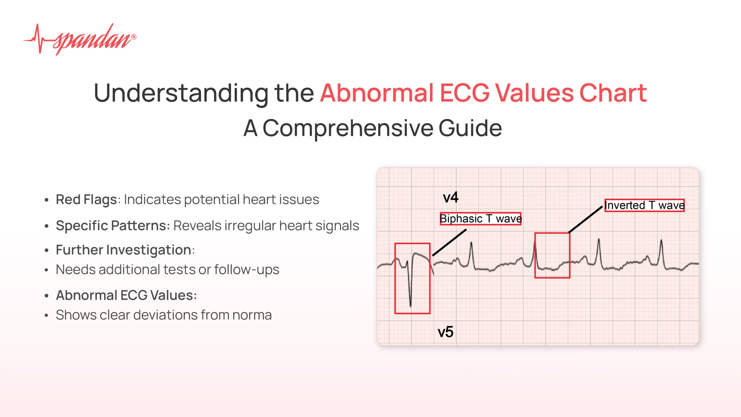
- Ischemia: Reduced blood flow to the heart muscle can cause ST segment depression or T wave inversion.
- Hypertrophy: Enlargement of the heart chambers can be detected by changes in the QRS complex.
- Conduction Abnormalities: Problems with the electrical pathways in the heart can cause prolonged PR interval or widened QRS complex.
- Electrolyte Imbalances: Abnormalities in potassium or calcium levels can affect the shape of the T wave and other ECG features.
Limitations of ECG Tests
While ECG tests are valuable diagnostic tools, they have some limitations:
- Snapshot in Time: A resting ECG only captures the heart’s electrical activity for a short period. It may not detect intermittent arrhythmias or other problems that occur sporadically.
- False Negatives: An ECG can be normal even if a person has underlying heart disease. This is especially true for conditions that are not present at rest.
- False Positives: An ECG can sometimes show abnormalities that are not actually related to a heart problem. This can lead to unnecessary testing and anxiety.
- Interpretation Variability: ECG interpretation can be subjective, and different healthcare professionals may have slightly different interpretations of the same ECG tracing.
- Not a Standalone Test: An ECG is usually used in conjunction with other diagnostic tests, such as blood tests, echocardiograms, or cardiac catheterization, to provide a more complete picture of the heart’s health.
Conclusion
The ECG test is a safe, non-invasive, and valuable tool for assessing the electrical activity of the heart. It plays a crucial role in diagnosing and managing a wide range of heart conditions. While ECG tests have some limitations, they remain an essential part of cardiovascular care. By understanding the purpose, procedure, and interpretation of ECG tests, patients can be better informed about their heart health and participate more actively in their medical care.


Leave a Reply