
“Advances in Diagnostic Imaging for Leukemia
Related Articles Advances in Diagnostic Imaging for Leukemia
- Alternative Therapies For Chronic Pain Management – Part 4
- Long-term Effects Of Chronic Illness On Children – Part 10
- Exercise And Physical Activity Guidelines For Chronic Illness Management – Part 4
- Yoga And Mindfulness Practices For Chronic Disease Patients – Part 4: Integrating Practices Into Daily Life And Maintaining Long-Term Adherence
- Holistic Approaches To Chronic Disease Prevention – Part 6: Cultivating Mindfulness And Stress Resilience
Introduction
With great enthusiasm, let’s explore interesting topics related to Advances in Diagnostic Imaging for Leukemia. Let’s knit interesting information and provide new insights to readers.
Advances in Diagnostic Imaging for Leukemia

Introduction
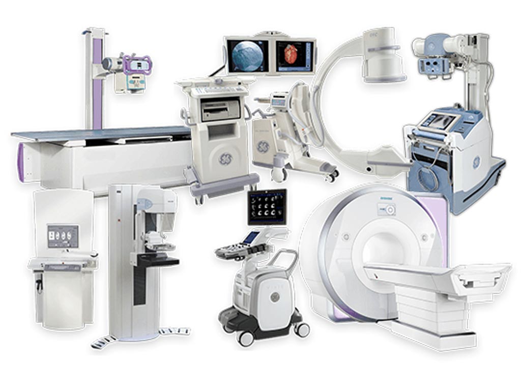
Leukemia, a group of cancers affecting the blood and bone marrow, poses a significant challenge in healthcare due to its diverse subtypes and complex pathogenesis. Accurate and timely diagnosis is crucial for effective treatment planning and improved patient outcomes. While traditional diagnostic methods like blood tests and bone marrow biopsies remain fundamental, advances in diagnostic imaging have revolutionized the way leukemia is detected, characterized, and monitored. This article explores the latest advancements in diagnostic imaging for leukemia, highlighting their clinical applications, benefits, and future directions.
The Role of Diagnostic Imaging in Leukemia Management
Diagnostic imaging plays a multifaceted role in leukemia management, encompassing:
-
Detection and Diagnosis: Imaging techniques can help identify the presence of leukemia by detecting abnormalities in the bone marrow, spleen, liver, and lymph nodes.
-
Subtype Characterization: Different leukemia subtypes exhibit distinct imaging features, aiding in accurate classification and risk stratification.
-
Disease Staging: Imaging is essential for determining the extent of disease involvement, including the presence of extramedullary disease (leukemia cells outside the bone marrow).
-
Treatment Monitoring: Imaging allows clinicians to assess treatment response by evaluating changes in tumor size, disease activity, and the presence of residual disease.
-
Relapse Detection: Imaging can detect early signs of relapse, enabling timely intervention and improved outcomes.
Conventional Imaging Techniques
-
X-ray: While not specific for leukemia, X-rays can reveal bone abnormalities, such as lytic lesions or fractures, which may be associated with certain leukemia subtypes.
-
Ultrasound: Ultrasound is primarily used to evaluate the spleen, liver, and lymph nodes for enlargement or abnormalities. It is non-invasive and readily available but has limited sensitivity for detecting subtle changes in the bone marrow.
-
Computed Tomography (CT): CT scans provide detailed cross-sectional images of the body, allowing for the assessment of organomegaly, lymphadenopathy, and extramedullary disease. CT is particularly useful for detecting leukemia-related complications, such as infections and bleeding.
-
Magnetic Resonance Imaging (MRI): MRI offers superior soft tissue contrast compared to CT, making it valuable for evaluating bone marrow infiltration, extramedullary disease, and central nervous system involvement. MRI is also preferred for imaging children with leukemia due to the absence of ionizing radiation.
Advanced Imaging Techniques
-
Positron Emission Tomography (PET): PET imaging utilizes radioactive tracers to detect metabolically active cells, including leukemia cells. 18F-fluorodeoxyglucose (FDG) PET/CT is the most commonly used PET technique in leukemia. It can help identify areas of increased glucose metabolism, indicating the presence of leukemia cells.
-
PET/MRI: PET/MRI combines the functional information from PET with the high soft tissue contrast of MRI, providing a comprehensive assessment of leukemia. PET/MRI is particularly useful for evaluating bone marrow involvement, extramedullary disease, and treatment response.
-
Diffusion-Weighted Imaging (DWI): DWI is an MRI technique that measures the movement of water molecules in tissues. It can help differentiate between benign and malignant lesions in the bone marrow and other organs. DWI is particularly useful for detecting early changes in the bone marrow during treatment.
-
Dynamic Contrast-Enhanced MRI (DCE-MRI): DCE-MRI assesses the blood supply to tissues by measuring the uptake and washout of contrast agents. It can help differentiate between active and inactive leukemia lesions and assess treatment response.
-
Molecular Imaging: Molecular imaging techniques use targeted probes to detect specific molecules or receptors on leukemia cells. These techniques can provide valuable information about the biology of leukemia and help personalize treatment.
Clinical Applications of Advanced Imaging in Leukemia
-
Diagnosis and Staging: PET/CT and PET/MRI can help diagnose leukemia by detecting areas of increased metabolic activity in the bone marrow, spleen, liver, and lymph nodes. These techniques can also help stage leukemia by determining the extent of disease involvement, including the presence of extramedullary disease.
-
Risk Stratification: Certain imaging features, such as the presence of extramedullary disease or high metabolic activity on PET/CT, may be associated with a higher risk of relapse or treatment failure. Imaging can help identify high-risk patients who may benefit from more intensive therapy.
-
Treatment Monitoring: PET/CT and PET/MRI can be used to monitor treatment response by evaluating changes in tumor size, metabolic activity, and the presence of residual disease. These techniques can help identify patients who are not responding to treatment and may need to switch to a different therapy.
-
Relapse Detection: Imaging can detect early signs of relapse, even before they are apparent on blood tests or bone marrow biopsies. Early detection of relapse allows for timely intervention and improved outcomes.
-
Minimal Residual Disease (MRD) Assessment: MRD refers to the presence of a small number of leukemia cells that remain after treatment. PET/CT and PET/MRI can help detect MRD in the bone marrow and other organs, providing valuable information about the risk of relapse.
-
Personalized Medicine: Molecular imaging techniques can help identify specific targets on leukemia cells, allowing for the development of personalized therapies that are tailored to the individual patient.
Benefits of Advanced Imaging Techniques
-
Improved Accuracy: Advanced imaging techniques, such as PET/CT and PET/MRI, offer superior accuracy compared to conventional imaging methods for detecting and staging leukemia.
-
Early Detection: Advanced imaging can detect leukemia at an earlier stage, allowing for timely intervention and improved outcomes.
-
Non-Invasive Assessment: Many advanced imaging techniques, such as PET/CT and MRI, are non-invasive and do not require bone marrow biopsies.
-
Comprehensive Assessment: Advanced imaging provides a comprehensive assessment of leukemia, including the bone marrow, spleen, liver, lymph nodes, and extramedullary sites.
-
Treatment Monitoring: Advanced imaging allows for the monitoring of treatment response and the detection of residual disease, enabling timely adjustments to therapy.
Limitations of Advanced Imaging Techniques
-
Cost: Advanced imaging techniques, such as PET/CT and PET/MRI, can be expensive.
-
Availability: Advanced imaging facilities may not be readily available in all healthcare settings.
-
Radiation Exposure: PET/CT involves exposure to ionizing radiation, although the dose is generally low.
-
False Positives: Advanced imaging techniques can sometimes produce false-positive results, leading to unnecessary investigations.
-
Interpretation Challenges: Interpretation of advanced imaging studies requires specialized expertise.
Future Directions
-
Development of Novel Tracers: Researchers are developing novel tracers for PET imaging that target specific molecules or receptors on leukemia cells, improving the accuracy and sensitivity of the technique.
-
Artificial Intelligence (AI): AI algorithms are being developed to analyze imaging data and improve the accuracy of diagnosis, staging, and treatment monitoring.
-
Radiomics: Radiomics involves extracting quantitative features from imaging data to predict treatment response and prognosis.
-
Multimodal Imaging: Combining different imaging modalities, such as PET/MRI and PET/CT, can provide a more comprehensive assessment of leukemia.
-
Image-Guided Therapy: Image-guided therapy involves using imaging to guide the delivery of treatment directly to leukemia cells, improving the effectiveness of therapy and reducing side effects.
Conclusion
Advances in diagnostic imaging have significantly improved the management of leukemia. Advanced imaging techniques, such as PET/CT, PET/MRI, and molecular imaging, offer superior accuracy, early detection, and comprehensive assessment compared to conventional imaging methods. These techniques play a crucial role in diagnosis, staging, risk stratification, treatment monitoring, relapse detection, and personalized medicine. While advanced imaging techniques have limitations, ongoing research and development are addressing these challenges and paving the way for even more effective and personalized approaches to leukemia management. As technology continues to evolve, diagnostic imaging will undoubtedly play an increasingly important role in improving the lives of patients with leukemia.


Leave a Reply